Brain Awareness Week: Five key terms behind the SEISMIC project
Every year, Brain Awareness Week brings together researchers, healthcare professionals and organisations around the world to raise awareness about the brain, neurological disorders and the importance of brain research.
To mark this year's Brain Awareness Week, we shared a series of "Brain Term of the Day" posts on our social media channels. Together, these five terms offer a glimpse into the clinical challenges and innovative technologies that lie at the heart of the SEISMIC project.
1. Intracerebral haemorrhage
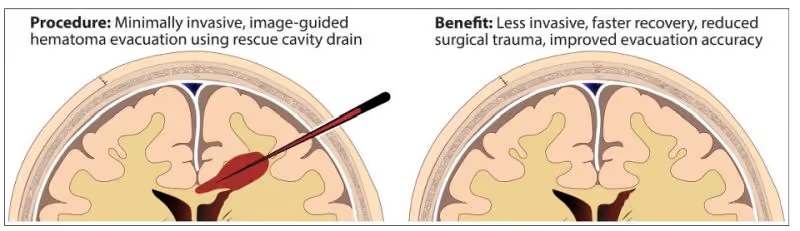
An intracerebral haemorrhage (ICH) is a type of stroke caused by bleeding deep within the brain tissue. It occurs when a blood vessel ruptures, allowing blood to accumulate inside the brain. The resulting pressure damages surrounding brain tissue, making ICH one of the deadliest forms of stroke.
Within SEISMIC, researchers are developing new image-guided approaches that could enable neurosurgeons to reach deep brain haemorrhages through a small opening in the skull. By combining real-time ultrasound with X-ray imaging, the project aims to support more precise, minimally invasive procedures that may improve patient outcomes.
2. Chronic subdural haematoma
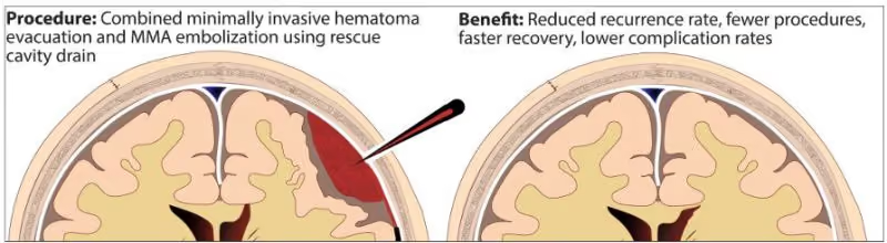
A chronic subdural haematoma (cSDH) is a collection of blood between the brain and the skull that often develops slowly after a minor head injury, particularly in older adults. Although current treatment is generally effective, the bleeding returns in a significant number of patients, often requiring additional surgery.
SEISMIC is investigating a new image-guided treatment strategy that combines drainage of the haematoma with embolisation of the middle meningeal artery. By addressing both the existing bleeding and its underlying cause, this approach has the potential to reduce recurrence and improve recovery. The project will also develop data-driven tools to identify patients at higher risk and support clinician training.
3. Cryoablation
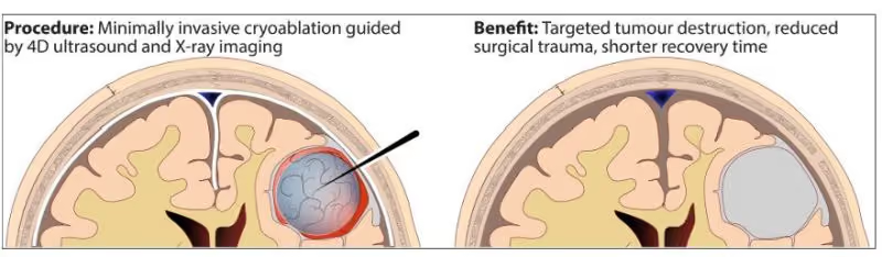
Cryoablation is a minimally invasive treatment that destroys tumour cells by exposing them to extremely low temperatures. Through carefully controlled cycles of freezing and thawing, tumour tissue can be targeted while preserving as much healthy tissue as possible.
Although cryoablation is already used successfully for several types of cancer, its application in brain tumours remains an emerging field. Within SEISMIC, researchers are developing image-guided cryoablation techniques specifically for neuro-oncology. By combining cryoablation with continuous intraoperative imaging, the project aims to replace large skull openings with a minimally invasive approach using a single burr hole, potentially expanding treatment options for patients with difficult-to-reach brain tumours.
4. Ultrasound for the brain
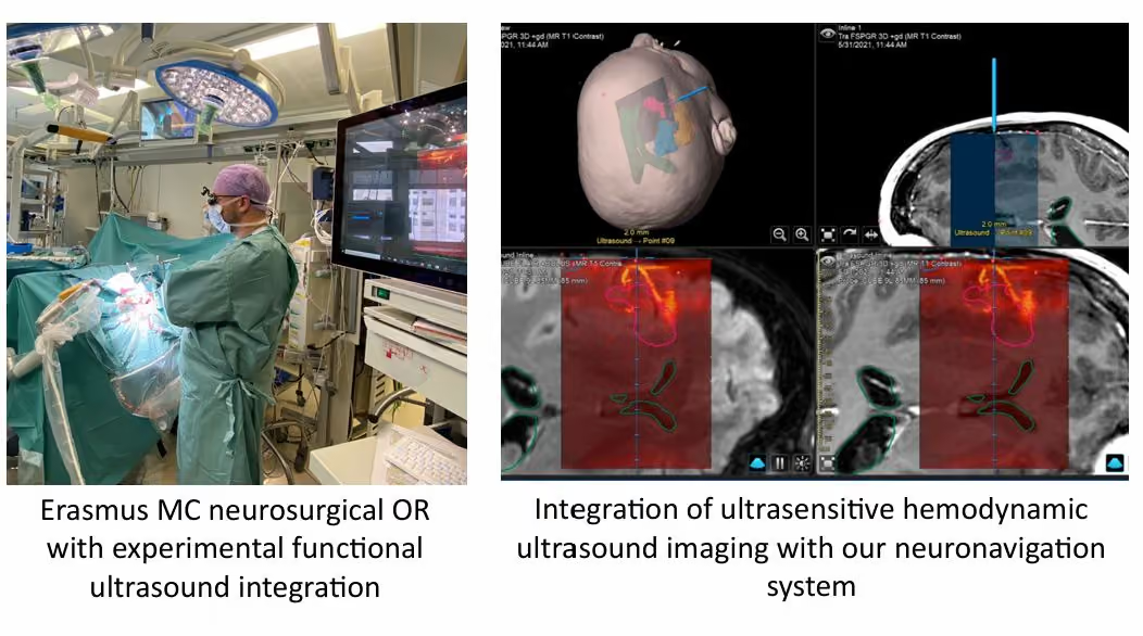
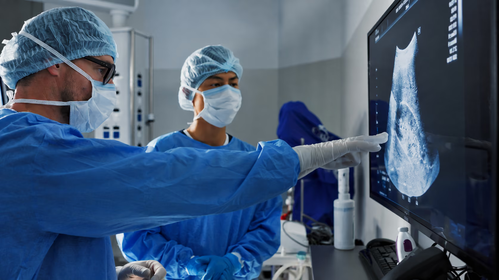
Unlike conventional imaging performed before surgery, intraoperative ultrasound allows surgeons to see the brain in real time while operating. Modern ultrasound technology can visualise brain structures, blood vessels and blood flow continuously without exposing patients to ionising radiation.
One of the major challenges during brain surgery is brain shift—the movement of brain tissue after the skull has been opened—which reduces the accuracy of preoperative scans. SEISMIC is developing an advanced 4D ultrasound system capable of continuously tracking these changes throughout surgery. Combined with X-ray imaging, this technology could provide surgeons with more accurate, real-time guidance during complex procedures.
5. Intraoperative X-ray
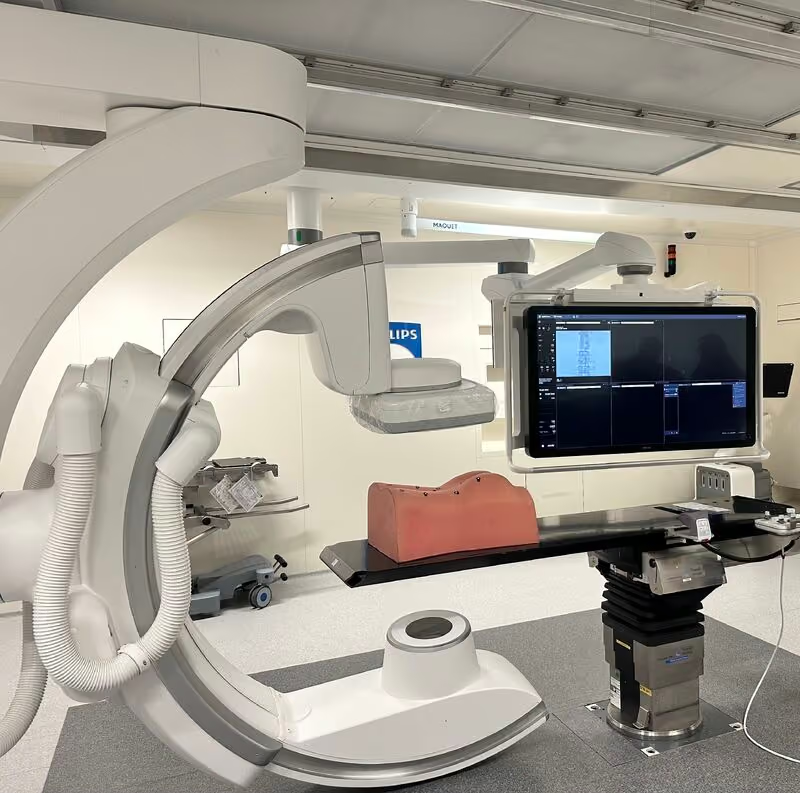
Intraoperative X-ray imaging provides high-resolution 2D and 3D images during surgery, allowing clinicians to visualise instruments and anatomical structures with great precision. However, acquiring these images can interrupt the surgical workflow and requires specialised equipment and expertise.
Within SEISMIC, researchers are working on a compact and flexible X-ray imaging solution that integrates seamlessly into the operating room. By combining the strengths of X-ray and advanced ultrasound imaging, the project aims to deliver richer real-time information while minimising disruption during surgery, ultimately supporting safer and more efficient procedures.
Looking ahead
These five topics represent just a small part of the scientific innovation taking place within SEISMIC. Over the coming years, our multidisciplinary consortium will continue developing technologies that bring together imaging, intervention and data science to make brain treatments safer, more precise and less invasive.
Brain Awareness Week reminds us that behind every technological innovation is a shared goal: improving the lives of patients affected by neurological disease. We look forward to sharing more updates, research highlights and educational content as the SEISMIC project progresses.