Advancing minimally invasive brain surgery through real-time imaging

Hemorrhagic Stroke
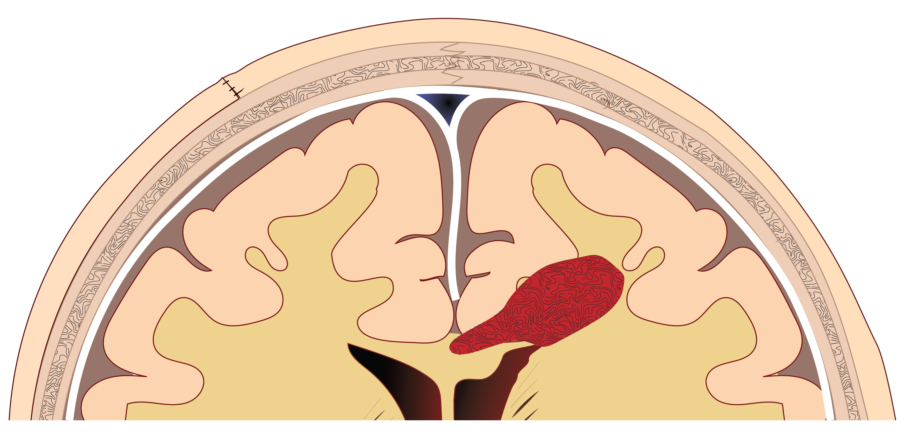
An intracerebral hemorrhage is a type of stroke caused by bleeding deep within the brain tissue.
- It happens when a blood vessel in the brain ruptures
- Blood accumulates, putting pressure on and damaging brain tissue
- It is the deadliest type of stroke and often leads to severe disability
- Rapid diagnosis and treatment are crucial to reduce brain injury
Subdural Bleeding
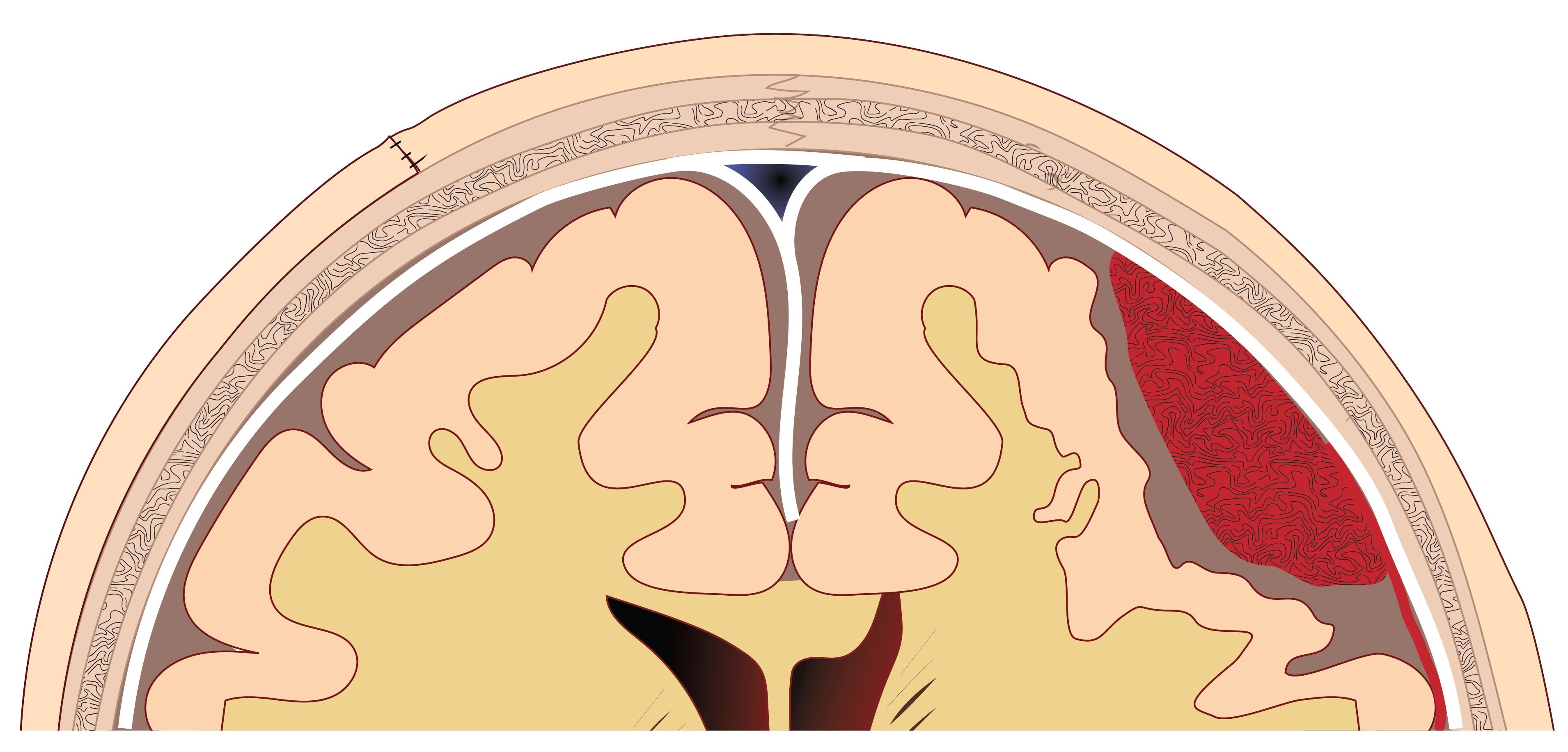
It is a chronic bleeding that occurs between the skull and the brain.
- It is common in older adults, and can develop slowly after a minor head injury
- Blood collects beneath the brain’s outer covering, putting pressure on the brain
- Standard treatment uses a small surgical hole to remove the blood
- Bleeding comes back in up to 30% of patients
- Recurrence leads to repeat surgeries, longer hospital stays, and more complications
Brain tumors
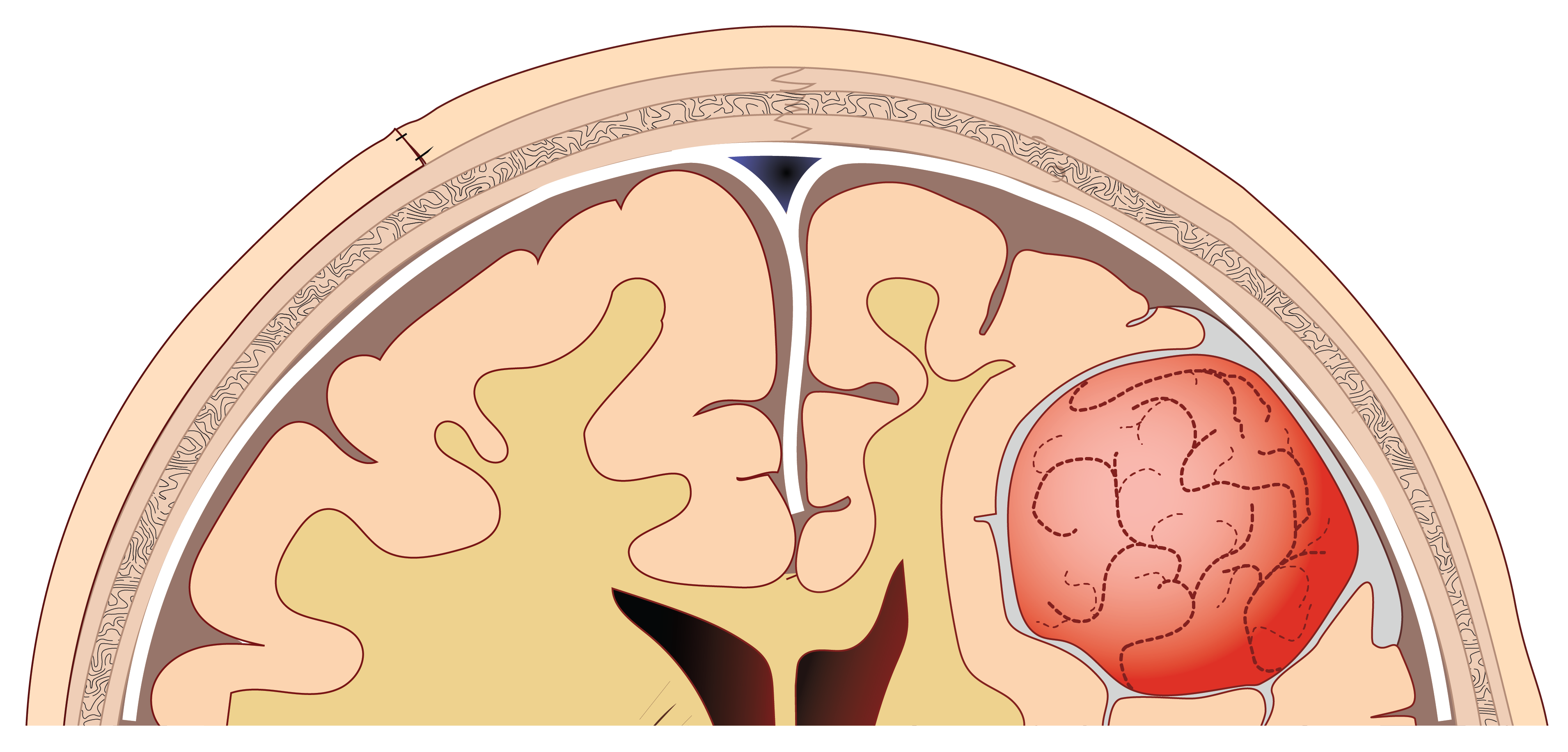
Abnormal growths of cells that develop in or around the brain. Key facts:
- Brain tumors can be benign or malignant
- Diagnosis is challenging because symptoms can develop slowly and gradually
- MRI scans and biopsies are commonly used, but determining the exact tumor type can still be difficult
- Treatment may include surgery and is delicate as the tumor is located near functional areas
Continuous 4D ultrasound
Ultrasound is a technique to see the brain in real time using sound waves. We will deliver the next generation of ultrasound image guidance. With this technology the surgeon can see deep brain structures and blood vessels continuously during surgery.
Intraoperative X-Ray
X-Ray is a medical imaging technique that uses a small dose of radiation to create pictures. The aim is to create a next generation robotic imaging platform capable of delivering high-performance intraoperative 2D&3D imaging. Together with the ultrasound, this information will improve surgical guidance.
Training simulator
A simulator is a realistic training system that lets healthcare professionals safely practice medical procedures without working on real patients. A high-fidelity surgical simulator will be developed to train clinicians in minimally invasive treatment of chronic bleeding.
Ultrasound & microbubbles
Microbubbles can safely be used to open the blood-brain barrier when ultrasound is applied. With this technique, surgeons will be able to determine tumor characteristics without invasive biopsies.
Cryoablation
Cyroablation is a technique that uses extremely cold temperatures to destroy tumors. We're developing image-guided cryoablation as a minimally invasive technique, where only a small needle needs to enter the brain.
How the technology supports surgery
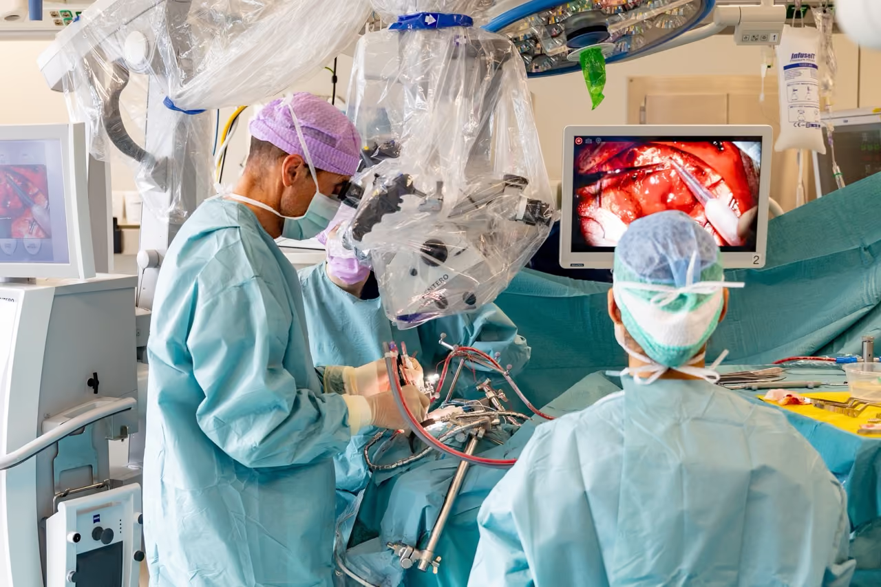
During brain surgery, surgeons operate in a highly constrained environment where visibility is limited. SEISMIC develops advanced imaging tools and minimally invasive treatment techniques that help surgeons:
- to identify critical structures
- expand treatment options for previously inoperable tumours/bleedings
- reduce complications
- ultimately improve both survival and quality of life for patients
Clinical validation in surgical practice
Our solution will be validated in 8 clinical studies. This ensures that our solutions are addressing real challenges and providing real benefit for the patients.
Brain tumour - treatment
Phase II study of image-guided cryoablation (extreme cold via a small skull opening) for low-accessibility brain tumours; safety, feasibility, early outcomes.
Brain tumour - diagnosis
Microbubble-enhanced focused ultrasound opens the blood-brain barrier for liquid biopsy, releasing brain-tumour biomarkers for minimally invasive diagnosis.
Training / simulation (SDH)
Development of a high-fidelity surgical simulator training module for minimally invasive chronic subdural haematoma procedures.
Subdural haematoma - drainage + MMA embolisation
Single image-guided procedure combining haematoma drainage with middle meningeal artery embolisation to reduce ~30% cSDH recurrence after surgery.
Subdural haematoma - outcomes
Development of quantitative and qualitative (patient-reported) outcome measures to standardise reporting in chronic subdural haematoma.
Subdural haematoma - registry
Multicentre NL registry investigating neurosurgical strategies and interventions guiding chronic subdural haematoma treatment and outcomes.
Intracerebral haemorrhage - approach validation
Anatomical validation and experimental models of minimally invasive surgical approaches to access and evacuate deep brain haemorrhage.
Intracerebral haemorrhage - treatment
SEISMIC ICH trial: image-guided minimally invasive drainage device to reach and evacuate deep intracerebral haemorrhage via a small skull opening.
Project roadmap
The technology is developed and evaluated in close collaboration with neurosurgeons. By testing the imaging approach in clinically relevant scenarios, researchers can assess how well the system visualises brain structures, supports surgical orientation and fits within existing surgical workflows. This ensures that the technology addresses real challenges faced during brain surgery.
December 2026
Key clinical and technical requirements defined
December 2027
Stakeholder advisory group established
January 2028
All clinical studies ready to start
Baseline operating room performance established
July 2028
First imaging prototypes deployed in hospitals
January 2029
Clinical studies reach mid-point recruitment target
January 2030
Assessment of clinical and healthcare value completed
January 2031
Final recommendations for future imaging systems delivered
Clinical studies completed